- Details
- ICNA
- News
- Hits: 796
Data presented at the 2015 American Epilepsy Society Annual Meeting in Philadelphia shows that sensitivity of the Brain Sentinel™ system for detection of GTCS is comparable to epileptologists reviewing vEEG recordings.
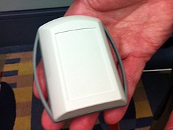
The system, which collects continuous surface electromyography (sEMG) and audio data via a device strapped to the bicep, is currently under review by the FDA.
The device algorithm continuously compares recorded surface EMG signals to the baseline sample of muscle activity. (GTC seizures are signalled with sustained activation of multiple frequency bands [30 to 40 Hz, 130 to 240 Hz, 300 to 400 Hz] of EMG activity.)
Quantitative review of sEMG may be a useful tool for defining motor components of epileptic and nonepileptic motor recruitment during seizures.
In this phase III double-blind controlled trial over 7800 hours of sEMG and vEEG data was reviewed for the 136 participants admitted to 11 epilepsy monitoring units in the U.S. Sensitivity to identify GTCS, when compared to vEEG review, was 100% CI (85-100), with false positive GTCS detection occurring at a rate of 1.4 false positives per 24 hours.
The device was able to alert patients of GTCS an average of 14 +/- 5 seconds after onset.
Neurologists may use this data to identify components of motor manifestation during GTCS or other times of interest reported by a patient. The device will not interfere with activities of daily living, and free patients of the stigma of epileptic seizure monitoring devices.
Reference:
Cavazos JE, Girouard M, Halford J, et al. Abstract 3.088. Automated EMG based Seizure Detection and Quantification for the Home and the Epilepsy Monitoring Unit: A Prospective Multicenter Study. Presented at: American Epilepsy Society Annual Meeting; Dec. 4-8, 2015; Philadelphia.
Disclaimer: The International Child Neurology Association (ICNA) or the American Epilepsy Society (AES) do not endorse or recommend any particular device or technology.
Read More

- Details
- ICNA
- News
- Hits: 869
Results from open-label Expanded Access treatment programs from 16 centres presented at the 2015 American Epilepsy Society Annual Meeting in Philadelphia, show that Epidiolex, a pharmaceutical-grade, purified form of cannabidiol, is generally well-tolerated and is a promising treatment for treatment-resistant epilepsies.
Epidiolex, manufactured by GW Pharma, is a highly-standardized, plant-derived form of cannabidiol (CBD), the most abundant non-psychoactive cannabinoid derived from the cannabis plant. Anecdotal reports and animal studies in multiple species and models have shown anticonvulsant efficacy for cannabidiol in intractable epilepsies including Dravet syndrome and Lennox-Gastaut syndrome.
For the current study by Devinsky et al, children and young adults with treatment-resistant epilepsy were enrolled in a 12-week prospective observational study. During a 4-week baseline period, parents and/or caregivers of patients kept prospective seizure diaries of all countable motor seizure types.
During the 12 weeks of CBD therapy, patients received a gradually increasing dose from 2-5 mg/kg/day until intolerance occurred or a maximum dose of 25 mg/kg/day was reached. During the treatment, patients were followed up every 2 to 4 weeks with complete blood count, liver, renal function, and antiepileptic drug levels done at baseline and at 4, 8, and 12 weeks of cannabidiol therapy.
261 patients (52% male; average age 11.8 years (range = 4 mo.-41 years old; average weight = 38 kg; average concomitant AEDs = 3) received 12 weeks of CBD therapy with data available at the last group collection. 44 of these patients had Dravet syndrome and 39 had a diagnosis of Lennox-Gastaut syndrome.
Following 3 months of CBD therapy, the median overall seizure frequency reduced by 45.1% in all patients and 62.7% in those with Dravet syndrome. In patients with Lennox-Gastaut syndrome there as a 71.7% median reduction of atonic seizures from baseline. Overall, 47% of all patients had at least a 50% reduction in seizures. At 3 months, seizure freedom occurred in 9% of patients and 13% of those with Dravet syndrome. Patients taking Clobazam had a higher rate of treatment response (57%) compared to those not on Clobazam co-therapy (39%).
Safety data from 313 patients from 16 sites was available. Twelve percent (36) of patients withdrew due to lack of efficacy. Adverse events occurred in more than 10% of patients, including somnolence (23%), diarrhoea (23%), fatigue (17%), decreased appetite (17%), convulsions (17%), and vomiting (10%).
Adverse events in 4% of patients led to discontinuation of CBD therapy. Serious adverse events were reported in 34% (106) of patients, including 7 deaths that were not considered to be treatment-related. Sixteen patients (5%) had serious adverse events considered to be treatment-related, including altered liver enzymes (4 patients [pts]; all were also on valproate and clobazam), status epilepticus/convulsion (4 pts), diarrhoea (4 pts), decreased weight (3 pts), thrombocytopenia (1 pt), and others.
These results are however from an uncontrolled study, and further study is needed to confirm the the safety and efficacy of Cannabidiol. Epidiolex is now being investigated in randomized controlled studies in DS and LGS.
Oldham M et al in related study looked at the the long-term efficacy of Epidiolex in 25 patients enrolled in the Expanded Access program at the University of California San Francisco and showed a greater than 50% reduction in seizure frequency in more than one-third of patients at 3-months following CBD treatment. The seizure control was maintained by 40% of patients for the 12-month duration.
25 patients (52% female) aged 1 to 17 years were enrolled in the program between January 2014 and December 2014. Twenty-eight percent of patients had Dravet syndrome, and epilepsy with myoclonic absences (EMA), CDKL5, and Lennox-Gastaut syndrome accounted for 16% each. Therapy period and treatment amounts followed the same protocols as in the Devinsky et al study. Median percent reduction in seizure frequency was assessed at 3 and 12 months, with treatment response considered ≥50% seizure reduction.
At 3-month follow-up, 8 patients (32%) had responded to CBD; 3 were seizure-free and 5 had a ≥50% seizure reduction. At 12-month follow-up, 10 patients (40%) had a ≥50% seizure reduction; 1 patient remained seizure-free. Twelve patients discontinued CBD due to lack of efficacy, and 1 patient due to increase in seizure frequency related to CBD.
Epidiolex is an oral formulation of pure plant-derived CBD. GW Pharmaceuticals plc is undertaking a formal development program for Epidiolex in severe, drug-resistant childhood epilepsy syndromes which include two pivotal Phase 3 trials of Epidiolex in Dravet syndrome and two pivotal Phase 3 trials of Epidiolex in Lennox-Gastaut syndrome. A Phase 3 trial in Tuberous Sclerosis Complex is due to commence shortly.
Reference
Devinsky O, Thiele E, Laux L, et al. Abstract 3.397. Efficacy and Safety of Epidiolex (Cannabidiol) in Children and Young Adults with Treatment-Resistant Epilepsy: Update from the Expanded Access Program. Presented at: American Epilepsy Society Annual Meeting; Dec. 4-8, 2015; Philadelphia.
Oldham M, Sullivan J, Singhal N, Tilton N, Cilio M. Abstract 2.296. Long-term efficacy and tolerability of add-on cannabidiol for drug-resistant pediatric epilepsies. Presented at: American Epilepsy Society Annual Meeting; Dec. 4-8, 2015; Philadelphia.
Source: AES
Read More

- Details
- ICNA
- News
- Hits: 839
In data was presented at the 2015 American Epilepsy Society Annual Meeting, Candace Myers, PhD, of the University of Washington, and colleagues have shown that targeted high throughput resequencing of genes in which a de novo mutation was previously been identified can help expand the phenotypic spectrum associated with these genes.
They sought to expand on their previous study where they identified 329 de novo mutations in 305 genes when 264 trios (affected child and unaffected parents).
For their current study the researchers performed targeted capture and high-throughput resequencing of 27 genes in which a de novo mutation was identified in one or more proband with Infantile Spasms (IS) or Lennox-Gastaut syndrome (LGS) in our prior study. 537 patients with diverse Epileptic Encephalopathy phenotypes were screened.
They were able to confirm the role of at least 7 additional genes in the genetic etiology of EE and expand the phenotypic spectrum associated with these genes beyond IS and LGS in which they were first discovered.
Among the genes identified there were recurrent and novel mutations in ALG13, CACNA1A, DNM1, GABRB3, GNAO1, IQSEC2, and SLC1A2 highlighting the importance of these genes in Epileptic Encephalopathies. Interestingly, 44% of the pathogenic variants identified in this study were recurrent mutations.
In addition they also identified a parent with a mosaic germline mutation in the case of two independent families with multiple affected individuals. GABRB3 accounted for the majority of pathogenic variants (n=6/537), accounting for ~1% of our cohort.
Citation: Myers C, McMahon J, Schneider A, et al. Abstract 1.315. Gene discovery in epileptic encephalopathies through targeted resequencing of candidate genes. Presented at: American Epilepsy Society Annual Meeting. Dec 4-8; Philadelphia.
Read More

- Details
- ICNA
- News
- Hits: 847
According to research presented at the 2015 American Epilepsy Society annual meeting in Philadelphia, dysfunction of brain circuits in pediatric patients with focal epilepsy results from a significant reduction in Circadian Locomotor Output Cycles Kaput (Clock) expression.
The researchers including Judy Liu MD PhD and her colleagues at the Children’s National Medical Center in Washington studied patients with focal epilepsy enrolled under their epilepsy surgery program. They used high-resolution 3t magnetic resonance imaging to determine epileptogenic foci and collected samples directly from the operating room for transcriptome analysis performed by microarray using Illumina® Gene Expression Bead Chip Array technology.
Studies in mice were also conducted for histological and gene expression analyses, whole cell patch-clamp electrophysiology, and pentylenetetrazole (ptz) seizure induction.
They found that compared with normal brain tissue the epileptogenic tissue showed a significant reduction of Circadian Locomotor Output Cycles Kaput (Clock) expression in 20 out of the 25 patients. Patients with decreased Clock included those with focal cortical dysplasias, tuberous sclerosis complex, Sturge-Weber syndrome, and Rasmussen's encephalitis.
The protein encoded by the Clock gene plays a central role in the regulation of circadian rhythms. Polymorphisms in this gene may be associated with behavioral changes, obesity, and metabolic syndrome. Two transcripts encoding the same protein have been found for this gene.
Deletion of the circadian clock in excitatory neurons in mice was associated with loss of spines in the apical dendrite and primary branches, a phenotype also seen in epileptogenic tissue of human pyramidal neurons.
These results point towards the key role of circadian genes in the pathogenesis of focal epilepsy in children and provide a link between the sleep-wake cycle and seizure threshold.
In addition it opens up the prospects of the circadian pathway being a promising target for therapeutic intervention.
Read More
- Details
- ICNA
- News
- Hits: 859
Research presented at the 2015 American Epilepsy Society Annual Meeting in Philadelphia shows that early detection of cognitive difficulties using a brief computerized cognitive screening may help improve delays in intervention. Cognitive problems and behavioural problems in epilepsy are frequent.
Cognitive difficulties in children with epilepsy affect different domains, including memory, language, and executive function that occur during critical periods of development. However screening tools currently available do not adequately identify these children, resulting in a delay in intervention.
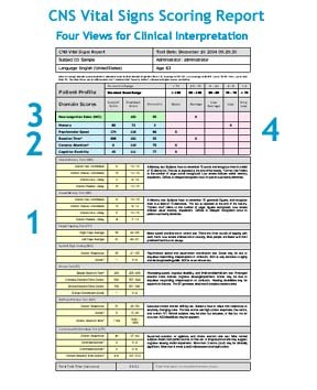
In their study Megan E. Bone, of the University of Pittsburgh School of Medicine, and colleagues evaluated the feasibility of the brief CNS Vital Signs (CNSVS) computerized cognitive battery for the detection of comorbidities in children with new-onset epilepsy.
They studied 33 patients (17 male; 26 generalized epilepsy, 7 focal epilepsy) aged 8-17 years with new-onset epilepsy and no previous antiepileptic drug treatment. Patients completed the CNSVS, which is completed in approximately 30 minutes, and parents completed the Strength and Difficulties Questionnaire, at two subsequent intervals (2-12 months = T2, 12-18 months= T3) during routine clinical appointments. Baseline scores were compared to subsequent scores using the Reliable Change Index (RCI).
All participants completed at least one follow up testing (mean follow-up time = 5.5 mo.), and 16 completed a third testing (mean total follow-up time = 14.5 mo.). Between baseline and first follow-up, 85% of patients had clinically significant changes in one of more cognitive domains including memory, psychomotor speed, reaction time, complex attention, and cognitive flexibility.
There was no apparent relationship between change in cognitive performance and seizure medication, epilepsy type, and seizure control. Composite memory, cognitive flexibility, reaction time, and complex attention showed the most change at both follow-up intervals, while no significant changes were observed for psychomotor speed.
In patients who improved or declined, change occurred at the first follow-up and remained stable thereafter. Parental concerns did not necessarily align with CNSVS score changes.
Citation: Bone ME, Triplett R, Rubin P, Asato MR. Abstract 1.293. Brief computerized screening detects cognitive changes in children with epilepsy. Presented at: American Epilepsy Society Annual Meeting; Dec 4-8, 2015; Philadelphia.
CNS Vital Signs (CNSVS) computerized cognitive battery
Read More
- Gene therapy against intractable epilepsy: promising proof of concept study
- De novo mutations in congenital heart disease and neurodevelopmental anomalies
- Decanoic acid, not ketones, behind therapeutic effect of MCT ketogenic diet
- Spiritam (Levetiracetam) world's first 3D printed drug gets FDA approval