- Details
- ICNA
- News
- Hits: 653
In pioneering research published in the Jan 18 issue of Neurology Pegoraro et al reports the identification of genetic modifiers of muscular dystrophy in humans for the first time.
Duchenne muscular dystrophy is a severe X-linked disease caused by mutations in the DMD gene that lead to nearly complete loss of dystrophin in skeletal and cardiac muscle.
In pioneering research published in the Jan 18 issue of Neurology Pegoraro et al reports the identification of genetic modifiers of muscular dystrophy in humans for the first time.The authors used informatics approaches, based on their earlier studies in normal subjects and from comparisons of rapid vs slow progression in DMD, to select a limited set of 29 candidate polymorphisms for analysis. One of the polymorphisms is a previously known single base variant in the promoter region of SPP1, and the authors provide evidence that the G allele of rs28357094 (observed in 35% of cases) is associated with more severe disease, as measured by either time to continuous wheelchair use or grip strength in 2 independent cohorts. The severe allele is present in about a third of all people, so this single allele could account for a substantial portion of the variability in disease progression.
In an accompanying editorial Drs Stanley F. Nelson and Robert C. Griggs point out that the presence of the G allele may provide a basis for stratifying patients entering clinical trials according to the anticipated rate of disease progression. This may improve the power of the trial by reducing interindividual heterogeneity, thereby permitting clinicians to select the relatively rapidly progressing subjects who may respond better to therapy.
In addition, the SPP1 variants associated with disease severity in DMD suggest the possibility of osteopontin as a rational therapeutic target.Osteopontin is a cytokine that promotes immune cell migration and survival; high tissue and circulating levels of osteopontin have been identified in a variety of muscular dystrophies, various inflammatory disorders, and cancer.
Studies of osteopontin knockout models have shown lessened severity and fibrosis in the mdx mouse. Interestingly however the G allele, associated with low osteopontin expression in mice and less disease seems to be associated with more severe disease in humans as shown by Pegoraro et al.
While hailing the findings of this study Drs Stanley F. Nelson and Robert C. Griggs also point out the importance of replicating these findings in larger cohort of DMD boys.It is indeed likely that all observed variations in DMD disease severity were not due to SPP1 variants.
With relatively inexpensive comprehensive sequencing of the human genome now made possible, this study should pave the way for more genome wide searches of disease modifiers as in the case of the recent identification of the IFRD1 variant in cystic fibrosis.
- SPP1 genotype is a determinant of disease severity in Duchenne muscular dystrophy E. Pegoraro, E.P. Hoffman, L. Piva, B.F. Gavassini, S. Cagnin, M. Ermani, L. Bello, G. Soraru, B. Pacchioni, M.D. Bonifati, G. Lanfranchi, C. Angelini, A. Kesari, I. Lee, H. Gordish-Dressman, J.M. Devaney, C.M. McDonald On behalf of the Cooperative International Neuromuscular Research Group Neurology 2011;76 219-226
- Predicting the severity of Duchenne muscular dystrophy: Implications for treatment Stanley F. Nelson and Robert C. Griggs Neurology 2011;76 208-20
Read More
- Details
- ICNA
- News
- Hits: 2255
Fever-induced refractory epileptic encephalopathy in school-age children (FIRES) is a recently described epileptic entity whose etiology remains unknown. Brain abnormalities shown by MRI are usually limited to mesial-temporal structures and do not account for the catastrophic neuropsychologic findings.
- Details
- ICNA
- News
- Hits: 817
Fever-induced refractory epileptic encephalopathy in school-age children (FIRES) is a recently described epileptic entity whose etiology remains unknown. Brain abnormalities shown by MRI are usually limited to mesial-temporal structures and do not account for the catastrophic neuropsychologic findings.
Research published in the January issue of The Journal of Nuclear Medicine shows that positron emission tomography (PET) scans can offer an evaluation of cognitive dysfunction of fever-induced refractory epileptic encephalopathy(FIRES), its evolution and further prognosis.
FIRES, a recently named condition, occurs in previously healthy children who, after a brief fever, experience acute seizures that are resistant to medication and last for several weeks. After the seizures stop, children are left with severe cognitive dysfunction, mainly involving language, memory and behavior.
The study, "18F-FDG PET Reveals Frontotemporal Dysfunction in Children with Fever-Induced Refractory Epileptic Encephalopathy," was conducted with eight patients diagnosed with FIRES. The patients were given a neuropsychologic evaluation, a brain MRI and an 18F-FDG PET scan. Severe cognitive dysfunction was noted, and while the MRI tests showed no abnormalities for the patients, the PET scans reported significant cognitive impairment.
Researchers compared the FIRES patients with a pseudo-control group of epilepsy patients with normal MRI and PET scan results. Using statistical parametric mapping, an objective approach to analyzing brain activity, the study exposed that the brain dysfunction was related to the epilepsy in the FIRES patients.
Reference
Mazzuca M, Jambaque I, Hertz-Pannier L, Bouilleret V, Archambaud F, Caviness V et al. (2011) 18F-FDG PET Reveals Frontotemporal Dysfunction in Children with Fever-Induced Refractory Epileptic Encephalopathy. J Nucl Med 52 (1):40-7. DOI: 10.2967/jnumed.110.077214 PMID: 21149491
Read More
- Details
- ICNA
- News
- Hits: 684
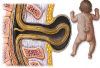 Women who suffer from epilepsy and take a common drug (carbamazepine) to treat the illness have a higher chance of having an infant with spina bifida compared with women not taking antiepileptic drugs, finds a study published on BMJ.com today.
Women who suffer from epilepsy and take a common drug (carbamazepine) to treat the illness have a higher chance of having an infant with spina bifida compared with women not taking antiepileptic drugs, finds a study published on BMJ.com today. Some women choose to terminate their pregnancy because their baby has this condition.
The researchers, led by Professor Lolkje de Jong-van den Berg from the University of Groningen in the Netherlands, carried out a review of all published studies to identify specific major malformations linked with carbamazepine use in the first three months of pregnancy.
Of the five identified indications in the literature, spina bifida was the only specific major congenital malformation significantly associated with exposure to carbamazepine monotherapy (spina bifida was 2.6 times more likely in infants of women who had taken carbamazepine compared with no antiepileptic drug, but the risk was smaller for carbamazepine than for valproic acid).
The authors did not conclude that carbamazepine is associated with other major malformations and say it is less risky than another frequently used antiepileptic drug, valproic acid.
They also stress that "although most antiepileptic drugs taken during pregnancy significantly increase the risk for one or more specific congenital malformations, the occurrence of these malformations is nevertheless rare ... most exposed pregnancies result in a baby without malformation." For carbamazepine taken in the first three months of pregnancy the overall risk of a major malformation was 3.3%.
Carbamazepine is one of the most commonly used anti-epilepsy drugs in Europe among women of reproductive age, says the paper.
The material was derived from EUROCAT - a database containing information from 19 registers of pregnancy outcomes with major congenital malformations in Europe from 1995 to 2005. The data relates to almost four million European births, of which over 98,000 involved a major malformation.
The authors say that their earlier study on valproic acid (published in the New England Journal of Medicine in 2010) concludes that women on valproic acid are six times more likely to have a pregnancy outcome with spina bifida and seven times more likely to have an outcome with hypospadias (a condition where a boy's urinary opening develops in the wrong part of the penis or in the scrotum) compared with women using other antiepileptic drug use. The research team therefore agrees with the recent recommendation from the American Academy of Neurology that women should avoid valproic acid in pregnancy if possible.
The authors conclude that although the overall risk of birth defects is low for women taking antiepileptic drugs, "the best option regarding antiepileptic drug treatment can be chosen only on an individual basis by the woman and neurologist before pregnancy, weighing the benefits of epilepsy control against the risk of teratogenicity."
In an accompanying editorial, Irena Nulman, Associate Professor of Paediatrics at the University of Toronto, says: "Of all the anticonvulsant drugs, "carbamazepine is associated with the lowest rate of morphological defects ... and should therefore be considered the drug of choice in pregnancy." She points out that, for many pregnant women, dicontinuing antiseizure drugs is not an option and that women should plan their pregnancy, receive evidence based prenatal counselling, and be given the safest antiepileptic drug.
Read More
- Details
- ICNA
- News
- Hits: 2249
Women who suffer from epilepsy and take a common drug (carbamazepine) to treat the illness have a higher chance of having an infant with spina bifida compared with women not taking antiepileptic drugs, finds a study published on BMJ.com today.