Researchers have shown that they were able to improve muscle function in Duchenne Muscular Dystrophy mice using in vivo gene editing techniques.
Duchenne muscular dystrophy (DMD) affects about 1 out of 5000 male births and caused by mutations in the dystrophin gene. Though DMD has been a target for gene therapy for a long time, progress has been very slow and attempts unsuccessful.The dystrophin gene has 79 sections, or exons, but can retain reasonable function even if a few exons in the middle are lost. Dystrophin works as long as its two ends are intact as in the case of Becker muscular dystrophy, in which frame shift mutations result in abnormal translation of the dystrophin gene.
New gene-editing techniques are now promising to make a substantial progress towards a treatment for Duchenne Muscular Dystrophy.Genome editing has the potential to restore expression of a modified dystrophin gene from the native locus to modulate disease progression by inserting the correct gene into the damaged cells. Removing one or more exons from the mutated transcript can produce an in-frame mRNA and a truncated, but still functional, protein.
An alternative treatment, using antisense oligonucleotides which are now in clinical trials works on the same principle of avoiding damaged exons, but instead of cutting them out of the DNA, they bind to the mutated exon, so that when the gene is then translated from the mature mRNA, it is “skipped” over, restoring the disrupted reading frame, which would still result in a largely functional protein.
Using the CrisprCas9 gene editing technique, researchers can cut the DNA of chromosomes at selected sites to remove or insert segments.
Three independent research groups have reported in the journal Science on 31 December 2015 that using the CrisprCas9 technique they were able to successfully treat mice with a defective dystrophin gene. All three groups had used a viral vector loaded with the DNA editing components to infect the muscle cells in DMD mouse and excise from the gene a defective exon. Without the defective exon, the muscle cells made a shortened truncated dystrophin protein which remained functional, giving the mice more strength.
The three teams were led by Charles A. Gersbach of Duke University, Eric N.Olson of the University of Texas Southwestern Medical Center and Amy J.Wagers of Harvard University.
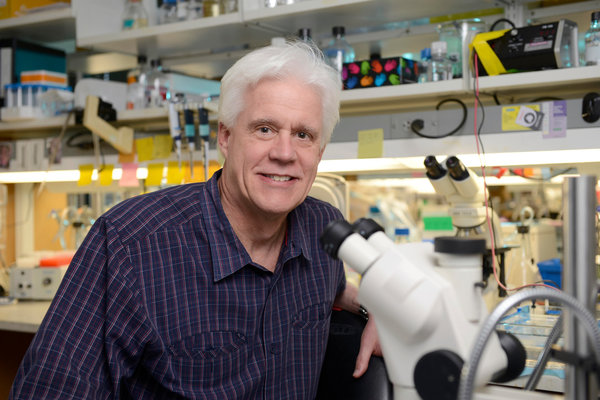 Eric N. OlsonIn 2014, the Olson group had reported on the successful editing out of the damaged 21st exon in a fertilized agg of the DMD mouse, thus causing an inheritable change to its genome. In this study they used adeno-associated virus-9 (AAV9) to deliver gene editing components to postnatal mdx mice. The gene editing components were directed to cut the two ends of the 21st exon. They tested different modes of AAV9 delivery including intra-peritoneal at postnatal day (P1), intra-muscular at P12, and retro-orbital at P18 and found that all three methods restored dystrophin protein expression in cardiac and skeletal muscle to varying degrees and expression increased from 3 to 12 weeks post-injection.
Postnatal gene editing also enhanced skeletal muscle function, measured by grip strength tests 4 weeks post-injection.The virus had successfully infected muscle cells throughout the mouse’s body, snipping out the exon from the dystrophin gene.The muscle cells repaired the DNA by joining the pieces of the cut chromosome and generated an effective dystrophin protein.
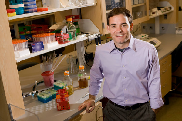 Charles A. GersbachGersbach's group had also reported earlier in 2015 that using the CrisprCas9 technique they were able to remove the 45th to 55th exons of the dystrophin gene from Duchenne
patient cell cultures. In their current study they also used the adeno-associated virus to deliver the CRISPR/Cas9 system to the mdx mouse model of DMD to remove the mutated exon 23 from the dystrophin gene. They used local and systemic delivery to adult mice and systemic delivery to neonatal mice.
Exon 23 deletion by CRISPR/Cas9 resulted in expression of the modified dystrophin gene, partial recovery of functional dystrophin protein in skeletal myofibers and cardiac muscle, improvement of muscle biochemistry, and significant enhancement of muscle force.
 Amy WagersWager's group on the other hand looked specifically at whether the genealtering virus could infect stem cells. In their study, they also used adeno-associated virus (AAV) of clustered regularly interspaced short palindromic repeats (CRISPR)-Cas9 endonucleases coupled with paired guide RNAs flanking the mutated Dmd exon23 which resulted in excision of intervening DNA and restored Dystrophin reading frame in myofibers, cardiomyocytes, and muscle stem cells following local or systemic delivery. AAV-Dmd CRISPR-treatment partially recovered muscle functional deficiencies and generated a pool of endogenously corrected myogenic precursors in mdx mouse muscle.
It is unclear whether CRISPR technique could be used to correct other types of mutations or how the viral vectors or the modified dystrophin gene may react with the human immune system.
Although gene therapy has been tried for Duchenne Muscular Dystophy in the past, there seems to be a real chance now of it being successful. All the three research groups have filed for patents and are optimistic that clinical trials could be launched in the near future.
Citations:
Long C, Amoasii L, Mireault AA, McAnally JR, Li H, Sanchez-Ortiz E et al. (2015) Postnatal genome editing partially restores dystrophin expression in a mouse model of muscular dystrophy.Science ():. DOI: 10.1126/science.aad5725 PMID: 26721683.
Tabebordbar M, Zhu K, Cheng JK, Chew WL, Widrick JJ, Yan WX et al. (2015) In vivo gene editing in dystrophic mouse muscle and muscle stem cells.Science ():. DOI: 10.1126/science.aad5177 PMID:26721686.
Nelson CE, Hakim CH, Ousterout DG, Thakore PI, Moreb EA, Rivera RM et al. (2015) In vivo genome editing improves muscle function in a mouse model of Duchenne muscular dystrophy.Science ():. DOI: 10.1126/science.aad5143 PMID: 26721684.
Cover image: Cross sections of muscle tissue from mice showing from left to right: normal healthy tissue, tissue with DMD and tissue after gene editing treatment source: Christopher Nelson, Duke University
Read More