- Neuroimages
- Leigh syndrome
Leigh syndrome
Updated
http://icnapedia.org/neuroimage/8772
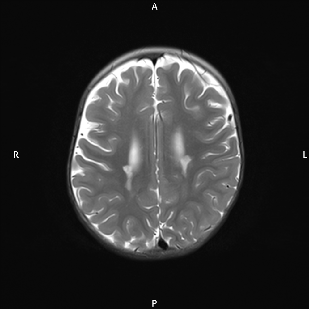
16 month old presented in type 2 respiratory failure secondary to apnoeas, investigations suggesting a possible central cause
T2 weighted images showing evidence of symmetric signal change within the cerebral peduncles where there is involvement of the corticospinal tracts and further signal change centrally, posterior pons but particularly in each inferior olivary nucleus and inferior cerebellar peduncles. There is also involvement of the nuclei of the tractus solitarius bilaterally. (These areas demonstrated restricted diffusion.) There is also some symmetric abnormal signal (which showed restricted diffusion) in the subthalamic nuclei also but no abnormality within either corpus striatum or thalamus. The brain is otherwise structurally normal. Myelination is normal for age at 16 months. The imaging features are highly suggestive of Leigh's Syndrome.
APA Style
Leigh syndrome. (n.d.). In ICNApedia. Retrieved August 24,2025 10:51:04 from http://icnapedia.org/neuroimage/8772
MLA Style
"Leigh syndrome." ICNApedia: The Child Neurology Knowledge Environment, Inc. May 12, 2020. Web. August 24,2025 10:51:04
AMA Style
ICNApedia contributors. Leigh syndrome. ICNApedia, The Child Neurology Knowledge Environment. May 12, 2020. Available at: http://icnapedia.org/neuroimage/8772.Accessed August 24,2025 10:51:04.


Add comment