-
Day 15 Post-Conception (p/c):
- Formation of the primitive streak of specialized neuroectoderm on the dorsal surface of the embryo.
-
Hensen’s Node:
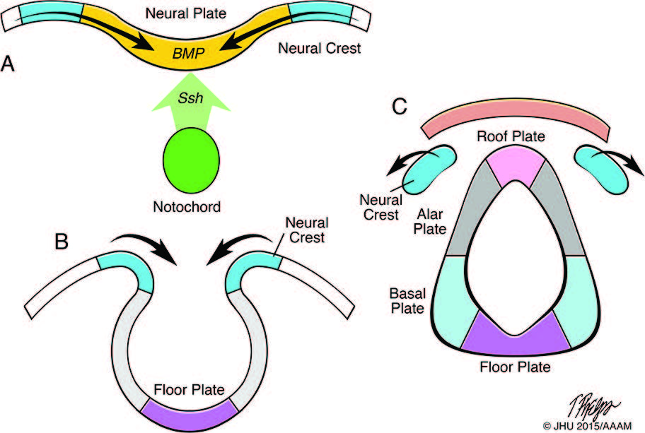
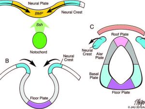 Development of the neural tube and neural crest. Fig. A shows the action of dorsalising signals from the ectoderm (e.g. bone morphogenetic proteins; BMP) and ventralising signals (e.g. sonic hedgehog, SHH) from the notochord on the developing neural plate. Fig. B shows folding of the edges of the neural plate to form the neural tube. Fig. C shows covering by the mesoderm and ectoderm over the closed neural tube, and separation of the neural crest tissues. The neural tube divided by the sulcus limitans into the dorsal alar and roof plates, and the ventral basal and floor plates. (Adapted from Ten Donkelaar, et al. Clinical Neuroembryology, 2nd edition, Springer 2014.)
- Small nodule at the rostral end of the neural plate.
- Directs development of the anterior neural tube.
-
Dorsal and Ventral Induction:
- Dorsal Induction:
- Formation and closure of the neural tube.
- Development of the three primary vesicles at the rostral end of the neural tube.
- Ventral Induction:
- Formation of cerebral hemispheres, eye vesicles, olfactory bulbs, pituitary glands, and part of the face.
- Includes primary and secondary neurulation.
- Dorsal Induction:
-
Primary Neurulation:
- Begins with the formation of the neural plate and tube.
- Neural plate formation starts on day 17 p/c and completes by day 18 p/c.
- Edges of the neural plate elevate and fold over to form the neural tube.
- Neural tube separates from the surface ectoderm by intervening mesenchyme.
- Closure starts on day 20 p/c at the level of the future rhombencephalon.
- Anterior neuropore closes by day 25 p/c and posterior neuropore by day 28 p/c.
-
Events During Neural Tube Closure:
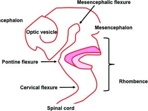 Closure of the anterior neural tube and folding into three vesicles the prosencephalon, mesencephalon, and rhombencephalon
- Disjunction: Neural tube separates from the cutaneous ectoderm which closes over the midline.
- Mesenchyme encircles the neural tube, forming the vertebral column, meninges, and muscle.
- Failure of neural tube closure can lead to lipomatous lesions associated with neural tube defects.
- Neural crest cells form, developing into dorsal root ganglia, cranial sensory and autonomic ganglia, and other tissues.
- Disjunction disturbances can result in congenital spinal lesions.
-
Secondary Neurulation:
- Starts with the closure of the posterior neuropore at the caudal eminence.
- Occurs in weeks 5 and 6 p/c, forming sacrococcygeal elements caudal to the closed posterior neuropore.
- Involves canalization with cyst formation and coalescence, forming the filum terminale and distal conus medullaris.
-
Ventral Induction (4 to 20 weeks p/c):
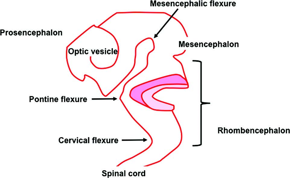
- Following closure of the anterior neuropore, three anterior neural tube vesicles (prosencephalon, mesencephalon, and rhombencephalon) form.
- Three major flexures (mesencephalic, pontine, and cervical) form in the anterior neural tube.
-
Patterning of the Neural Primordium:
- Regionalization by segmented cell differentiation across the developing neuroaxis.
- Controlled by spatial and temporal gene expression along rostrocaudal, dorsoventral, and mediolateral axes.
- Morphogenetic gradients of inductive signaling determine regional neural cell phenotypes.
- Neural primordium divided into segments or neuromeres, each with a floor, basal, alar, and roof plate.
- Special signaling centers, or secondary organizers, refine local neural identities along the neural tube.
-
Secondary Organizers:
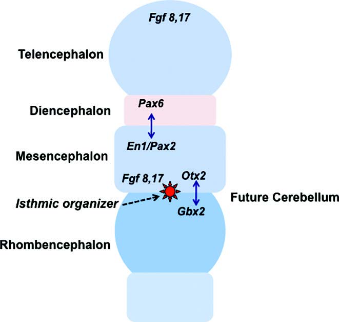
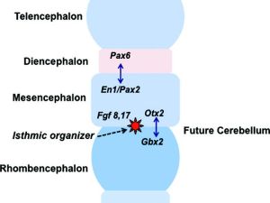 Patterning of neural territories in the anterior neural tube by organisers. Diagram showing the definition of the fundamental territories of the anterior neural tube by complex and dynamic effects of suppressor and permissive gene products. Note position of the isthmic organiser (IsO) at the mesencephalic-rhombencephalic junction, the location of the cerebellar anlage.
- Identified Locations:
- Anterior neural ridge (rostral edge of the neural plate).
- Zona limitans interthalamica (diencephalon).
- Isthmic organizer (midbrain-hindbrain junction).
- Functions:
- Responsible for graded expression of dorsalizing and ventralizing factors.
- Generate ventral motor and dorsal sensory cells of the neural tube.
- Dorsalizing Factors:
- Bone morphogenic protein (BMP) family produced by the non-neural ectoderm of the roof plate.
- Ventralizing Factors:
- Proteins expressed by the sonic hedgehog (SHH) gene in the prechordal and floor plates.
- Identified Locations:
Cite this: ICNApedia contributors.Neural tube development. ICNApedia, The Child Neurology Knowledge Environment. 28 June 2024. Available at: https://icnapedia.org/knowledgebase/articles/neural-tube-development Accessed 28 June 2024.