This is an old revision of the document!
Index
Quick yet systematic approach to MR imaging of brain lesions
In general, there are 4 MR sequences that will tell you 99% of what you need to know:
1. T2 FLAIR 2. T1 post-contrast 3. DWI 4. GRE/SWI/SWAN/T2*
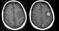
1. T2 FLAIR: “What is grossly abnormal?” anything abnormal = bright on T2 FLAIR 2. T1+gadolinium*: “Where is the BBB disrupted?”
Also:
-“What is the vascular composition?”
always compare T1+gad sequences to pure T1 without contrast (if also bright on pure T1, then BBB not necessarily disrupted)
 3. DWI*:
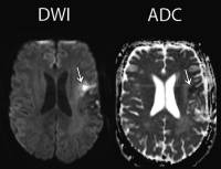
bright = abnormal = “diffusion-restricting” = could be hypercellularity, ischemia, demyelination, abscess, &c
the above applies only if the corresponding area is dark on ADC sequence (if also bright on ADC, then just T2 shine-through)
 4. GRE/SWI/SWAN/T2*: helps to distinguish nuances among entities on your differential.
in general, abnormal = dark (blood, vascularity, &c; abscesses tend to have dark rims on SWI)

