CLINICAL AND RADIOLOGICAL PROFILE OF FOCAL CORTICAL DYSPLASIA IN CHILDREN
Shikha Jain, Prateek Malik, Sangeetha Yoganathan, Karthik Muthusamy, Maya Mary Thomas
Christian Medical College, Vellore, India
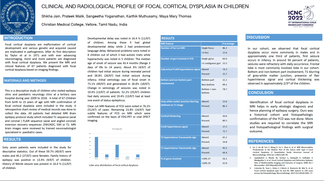
Focal cortical dysplasia are malformation of cortical development and various genetic and acquired caused are implicated in pathogenesis. This is a hospital based study of patients who visited epilepsy clinic at a tertiary care hospital during year 2009 to 2018. A total of 67 Children from birth to 15 years of age with MRI confirmation of focal cortical dysplasia were included in the study. Out of these 59.7% (40/67) were males and 40.2 (27/67) were females. Family history of epilepsy was positive in 13.4% (9/67) of children. Developmental delay was noted in 16.4 % (11/67) of children. Learning problems were noted in 18 children. The median age of onset of seizure was 43.4 months (Range 3 days of life to 14 years). About 6% (4/67) of children had initial seizure during neonatal period and 38.8% (26/67) had initial seizure during infancy. Initial semiology was of focal onset in 73.1% (49/67) and generalized in 26.8% (18/67). Change in semiology of seizures was noted in 19.4% (13/67) of patients. 52.2% (35/67) children had daily seizures. Clear cut MRI features of FCD were noted in 76.2% (51/67) of cases. Remaining 23.8% (16/67) had subtle features of FCD on MRI. The most common site of occurrence of focal cortical dysplasia was frontal lobe (50.7%). Second commonest site for occurrence of FCD was temporal lobe (31.3%). Only one patient had FCD in occipital lobe. Five patients (7.4%) had focal cortical dysplasias involving multiple lobes.
Keywords: focal cortical dysplasia, epilepsy
Institution: Christian Medical College, Vellore, India