A Case Of Vanishing White Matter Disease With Atypical Neuroimaging Presentation
OLGAY BILDIK, ELIF DIDINMEZ TASKIRDI, MELIS KOSE, NIHAL OLGAC DUNDAR, PINAR GENCPINAR
Objectives Vanishing white matter disease (VWMD) is a recessive neurodegenerative disease caused by eIF2B gene mutations. The disease is characterized by progressive loss of white matter in both hemispheres of the brain. An important feature of the disease is the worsening of clinical symptoms when exposed to physiological and environmental stress factors . We aimed to present our patient because of atypical localized brain Magnetic resonance imaging(MRI) results and different electromyography(EMG) results.
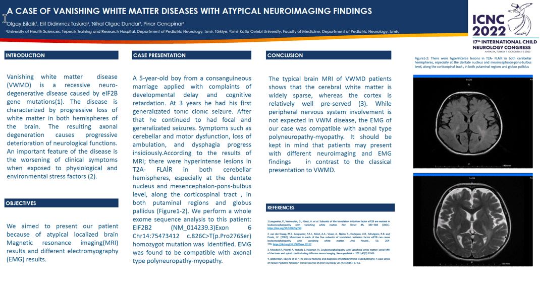
Case presentation A 5-year-old boy from a consanguineous marriage applied with complaints of developmental delay and cognitive retardation. At 3 years he had his first generalızated tonıc clonıc seizure. Symptoms such as cerebellar and motor dysfunction, loss of ambulation, and dysphagia progress insidiously. According to the results of MRI; there were hyperintense lesions in T2A- FLAİR in both cerebellar hemispheres, especially at the dentate nucleus and mesencephalon-pons-bulbus level. We perform a whole exome sequence analysis to this patient: EIF2B2 (NM_014239.3)Exon 6 Chr14:75473412 c.826C>T(p.Pro276Ser) homozygot mutation was identified. EMG was found to be compatible with axonal type polyneuropathy-myopathy.
Discussion The typical brain MRI of VWMD patients shows that the cerebral white matter is widely sparse, whereas the cortex is relatively well pre-served. In English literatüre 96% of EMG (both juvenile and late infantile metachromatic leukodystrophy forms) was abnormal with demyelinating sensorimotor neuropathy pattern, the EMG of our case was compatible with axonal type polyneuropathy-myopathy.It should be kept in mind that patients may present with different neuroimaging and EMG findings in contrast to the classical presentation of VWMD.
Keywords: vanishing white matter diseases, polyneuropathy
OLGAY BILDIK
University of Health Sciences, Tepecik Training and Research Hospital
Turkey
ELIF DIDINMEZ TASKIRDI
University of Health Sciences, Tepecik Training and Research Hospital
Turkey
MELIS KOSE
Izmir Katip Celebi University, Faculty of Medicine
Turkey
NIHAL OLGAC DUNDAR
Izmir Katip Celebi University, Faculty of Medicine
Turkey
PINAR GENCPINAR
Izmir Katip Celebi University, Faculty of Medicine
Turkey

OLGAY BILDIK
University of Health Sciences, Tepecik Training and Research Hospital Turkey
University of Health Sciences, Tepecik Training and Research Hospital Turkey