Posterior Ocular Structure Parameters by Optical Coherence Tomography Angiography in Pediatric Epilepsy patients
Pembe Gültutan, Pınar Nalçacıoğlu Memiş , Deniz Yılmaz, Mehmet İçöz, Ayşegül Neşe Çıtak Kurt
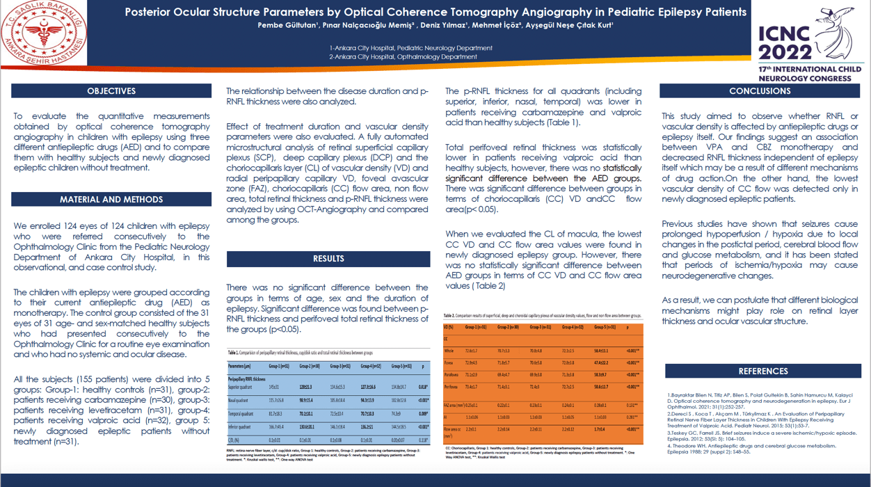
Objectives: To evaluate the quantitative measurements obtained by optical coherence tomography angiography in children with epilepsy using three different antiepileptic drugs (AED) and to compare them with healthy subjects and newly diagnosed epileptic children without treatment.
Methods: This observational, cross-sectional study included 155 patients divided into 5 groups: Group-1: healthy controls (n=31), group-2: patients receiving carbamazepine (n=30), group-3: patients receiving levetiracetam (n=31), group-4: patients receiving valproic acid (n=32), group 5: newly diagnosed epileptic patients without treatment (n=31). A fully automated microstructural analysis of the foveal avascular zone (FAZ), FAZ perimeter, foveal vessel density in a 300-µm area around the FAZ, nonflow area, flow index in superficial and deep vascular plexus, choriocapillary (CC) flow, vascular density, radial peripapillary capillary density was performed. Effect of treatment duration and vascular density parameters were also evaluated. Results: The mean age, gender distribution and the treatment duration were similar in all groups. There was statistically significant difference between the groups for vascular density of CC (VDC), CC flow area, and p-RNFL thickness. p-RNFL thickness was lower in groups 2 and 4. While the lowest VDC flow was in group-5, there was no significant difference between AED groups. Conclusion: In the present study carbamazepine and valproic acid decreased the p-RNFL thickness which was not observed in the levetiracetam group. While lowest vascular density of cc flow was detected in newly diagnosed epileptic patients, there was no difference between the treatment groups in terms of vascular density parameters.
Keywords: epilepsy,Optic coherence tomography angiography,peripapillary retina nerve fiber layer,peripapillary vascular density
Pembe Gültutan
Ankara City Hospital, Children's Hospital , Pediatric Neurology
Turkey
Pınar Nalçacıoğlu Memiş
Ankara City Hospital, Opthalmology
Turkey
Deniz Yılmaz
Ankara City Hospital, Children's Hospital , Pediatric Neurology
Turkey
Mehmet İçöz
Ankara City Hospital, Opthalmology
Turkey
Ayşegül Neşe Çıtak Kurt
Ankara City Hospital, Children's Hospital , Pediatric Neurology
Turkey
Pembe Gültutan
Ankara City Hospital, Children's Hospital , Pediatric Neurology Turkey
Ankara City Hospital, Children's Hospital , Pediatric Neurology Turkey