STURGE WEBER SYNDROME WITH PHAKOMATOSIS PIGMENTOVASCULARIS: A CASE REPORT
Aakanksha anand, Suvasini Sharma, dipti kapoor
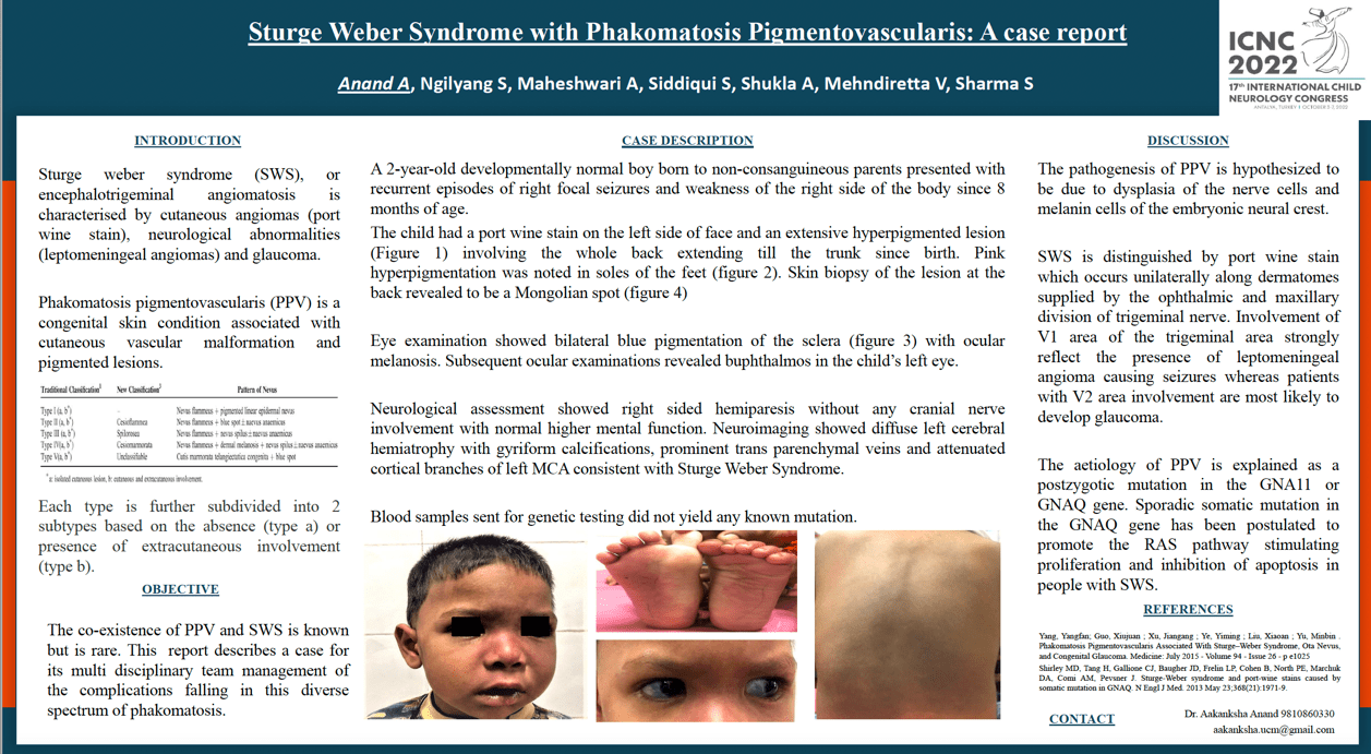
Sturge weber syndrome is a neurocutaneous syndrome characterised by cutaneous angiomas (portwine stain), neurological abnormalities (leptomeningeal angiomas) and glaucoma. Phakomatosis pigmentovascularis (PPV) is a congenital skin condition associated with cutaneous vascular malformation and dermal melanosis with or without ocular and systemic involvement. A 2-year-old developmentally normal boy born to non-consanguineous parents presented with recurrent right focal seizures and transient weakness of the right side of the body since 8 months of age. The child had a port wine stain on the left side of face and a large hyperpigmented lesion at the back since birth. Eye examination showed bilateral blue pigmentation of the sclera along with hyperpigmentation. Neurological assessment showed right sided hemiparesis without any cranial nerve involvement without disrupting higher mental function. Neuroimaging showed diffuse left cerebral hemiatrophy with gyriform calcifications, prominent transparenchymal veins and attenuated cortical branches of left middle cerebral artery consistent with Sturge Weber Syndrome. Electroencephalogram showed a normal study. Subsequent ocular examinations revealed buphthalmos in the child’s left eye. The etiology of PPV is explained as a postzygotic mutation in the GNA11 or GNAQ gene. Sporadic somatic mutation of the GNAQ gene has been recognized as the etiology for Sturge–Weber syndrome. Pediatric neurologists, geneticists and dysmorphologists need to develop an increased awareness of PPV and its association with various syndromes with systemic involvement.
Keywords: Sturge-weber, phakomatosis
Aakanksha anand
ESIC Medical College & Hospital
India
Suvasini Sharma
Lady Hardinge Medical College
India
dipti kapoor
Lady Hardinge Medical College
India

Aakanksha Anand
ESIC Medical College & Hospital India
ESIC Medical College & Hospital India