Neurodegeneration with brain iron accumulation in 18 Indian families: a case series highlighting phenotypic and genotypic diversity
Amita Moirangthem, Haseena Sait, Somya Srivastava, Manmohan Pandey, Deepak Ravichandran, Anju Shukla, Kausik Mandal, Deepti Saxena, Arya Shambhavi, Purvi Majethia, Lakshmi Priya Rao, Suvasini Sharma, Shubha Phadke
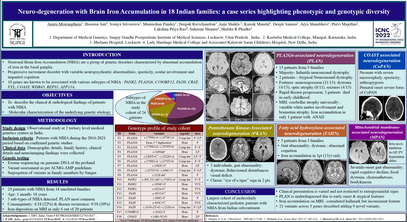
Objectives: To study the clinical, neuroimaging and genetic spectrum of disorders with neurodegeneration with brain iron accumulation (NBIA). Methods: All molecular proven cases of NBIA presenting in last 5 years at 2 tertiary care genetic centers were compiled. Demographic details, clinical and neuroimaging findings were collated. The probands were genotyped by exome sequencing. Results: We describe 24 individuals from 18 unrelated families with causative variants in 5 NBIA-associated genes. PLA2G6-associated neurodegeneration was the most common, observed in 13 individuals from 9 families. They presented mostly in infancy with neuroregression and hypotonia. A recurrent pathogenic variant in COASY was observed in a neonate with prenatal-onset severe neurodegeneration. Pathogenic bi-allelic variants in PANK2, FA2H and C12ORF genes were observed in the rest and these individuals presented in late childhood and adolescence with gait ataxia and dystonia. Iron deposition on neuroimaging was seen in only 5/16 patients. The classical “eye of tiger” sign was observed in only 2 patients with PKAN whereas cerebellar atrophy was a universal feature across all subtypes. A total of 21 different causative variants were detected including a multiexonic duplication in PLA2G6. The variants c.1799G>A and c.2370T>G in PLA2G6 were observed in three unrelated families each. In-silico assessments of the 9 novel variants were also performed. No intra and interfamilial variability was observed. Conclusion: We present a comprehensive compilation of phenotypic and genotypic spectrum of NBIA subtypes from the Indian subcontinent. Clinical presentation of NBIA is varied and not restricted to extrapyramidal features or iron accumulation on neuroimaging.
Keywords: dystonia, neurodegeneration, iron deposition, basal ganglia, PLA2G6
Amita Moirangthem
Sanjay Gandhi Postgraduate Institute of Medical Sciences
India
Haseena Sait
Sanjay Gandhi Postgraduate Institute of Medical Sciences
India
Somya Srivastava
Sanjay Gandhi Postgraduate Institute of Medical Sciences
India
Manmohan Pandey
Sanjay Gandhi Postgraduate Institute of Medical Sciences
India
Deepak Ravichandran
Medanta Hospital
India
Anju Shukla
Kasturba Medical College, Manipal, Manipal Academy of Higher Education
India
Kausik Mandal
Sanjay Gandhi Postgraduate Institute of Medical Sciences
India
Deepti Saxena
Sanjay Gandhi Postgraduate Institute of Medical Sciences
India
Arya Shambhavi
Sanjay Gandhi Postgraduate Institute of Medical Sciences
India

Amita Moirangthem
Sanjay Gandhi Postgraduate Institute of Medical Sciences India
Sanjay Gandhi Postgraduate Institute of Medical Sciences India