Balamuthia Mandrillaris Granulomatous Amoebic Encephalitis: A Report Of Two Cases
Abstract: Balamuthia mandrillaris is a free-living amoeba found in soil and freshwater. Infection occurs through the skin or the respiratory tract, with cutaneous and CNS involvement occurring from haematogenous spread. Balamuthia amoebic encephalitis (BAE) presents with granulomas, necrosis and haemorrhage and is nearly always fatal.
Case 1
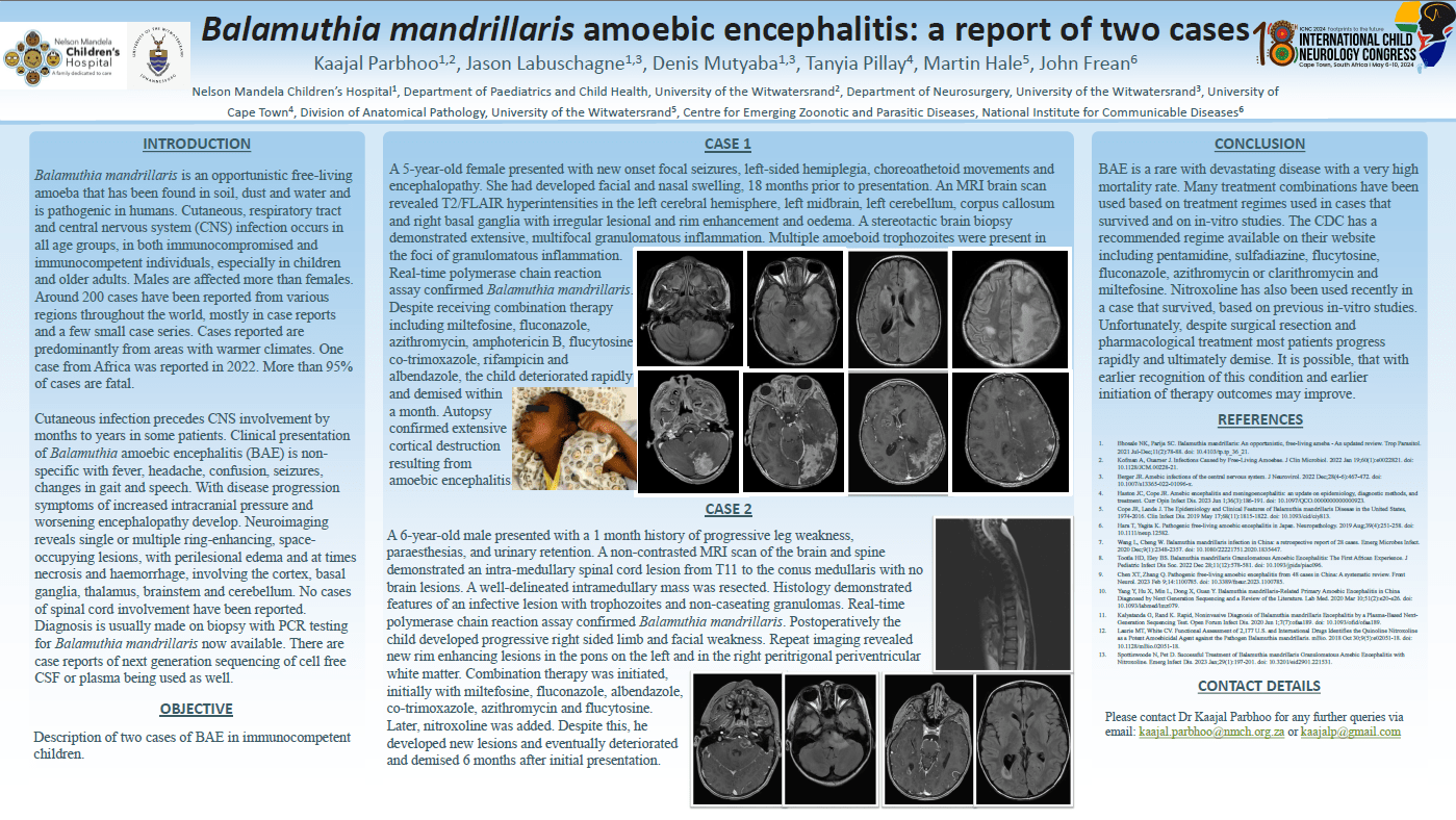
6-year-old male presented with a 1-month history of progressive leg weakness and urinary retention. MRI demonstrated an intramedullary spinal cord lesion. Histology demonstrated features of an infective lesion with non-caseating granulomas. A real-time polymerase chain reaction (PCR) assay confirmed Balamuthia mandrillaris. Initial brain MRI was normal, but he later he developed multiple brain lesions. Despite initially improving on combination therapy (miltefosine, fluconazole, albendazole, co-trimoxazole, azithromycin, flucytosine, nitroxoline), he progressed after a few months and demised.
Case 2
5-year-old female presented with an 18-month history of nasal swelling followed by a 1-month history of focal seizures, hemiplegia and encephalopathy. MRI scan demonstrated extensive T2/FLAIR-hyperintensities involving the cortex, brainstem and basal ganglia with rim enhancing areas. Stereotactic biopsy revealed extensive, multifocal granulomatous inflammation on histology with multiple amoeboid trophozoites. A real-time PCR assay confirmed Balamuthia mandrillaris. Despite receiving combination therapy (miltefosine, fluconazole, azitrhomycin, amphotericin B, flucytosine, co-trimoxazole, rifampicin, albendazole) the child rapidly deteriorated and demised. Autopsy confirmed extensive cortical destruction resulting from amoebic encephalitis.
Balamuthia infections are rare, with only about 200 cases reported worldwide, and 1 case reported in Africa. Balamuthia encephalitis should be considered in granulomatous CNS infections. Increased awareness may lead to earlier diagnosis and initiation of treatment with improved outcomes.
Kaajal Parbhoo
Paediatric Neurologist
Paediatric Neurology
University of the Witwatersrand
Johannesburg, South Africa
Jason Labuschagne
Neurosurgeon, Neurosurgery
University of the Witwatersrand
Johannesburg, South Africa
Denis Mutyaba
Neurosurgeon
Neurosurgery
University of the Witwatersrand
Johannesburg
South Africa
Tanyia Pillay
Radiologist
Radiology
University of Cape Town
Cape Town, South Africa
Martin Hale
Anatomical Pathologist
Anatomical Pathology
University of the Witwatersrand
Johannesburg, South Africa
John Frean
Head of Parasitology Reference Laboratory
Centre for Emerging Zoonotic and Parasitic Diseases
National Institute for Communicable Diseases, Division of the National Health Laboratory Service