Fetal Neurological Malformations Where Doppler Solved The Riddle: A Series Of Three Independent Cases
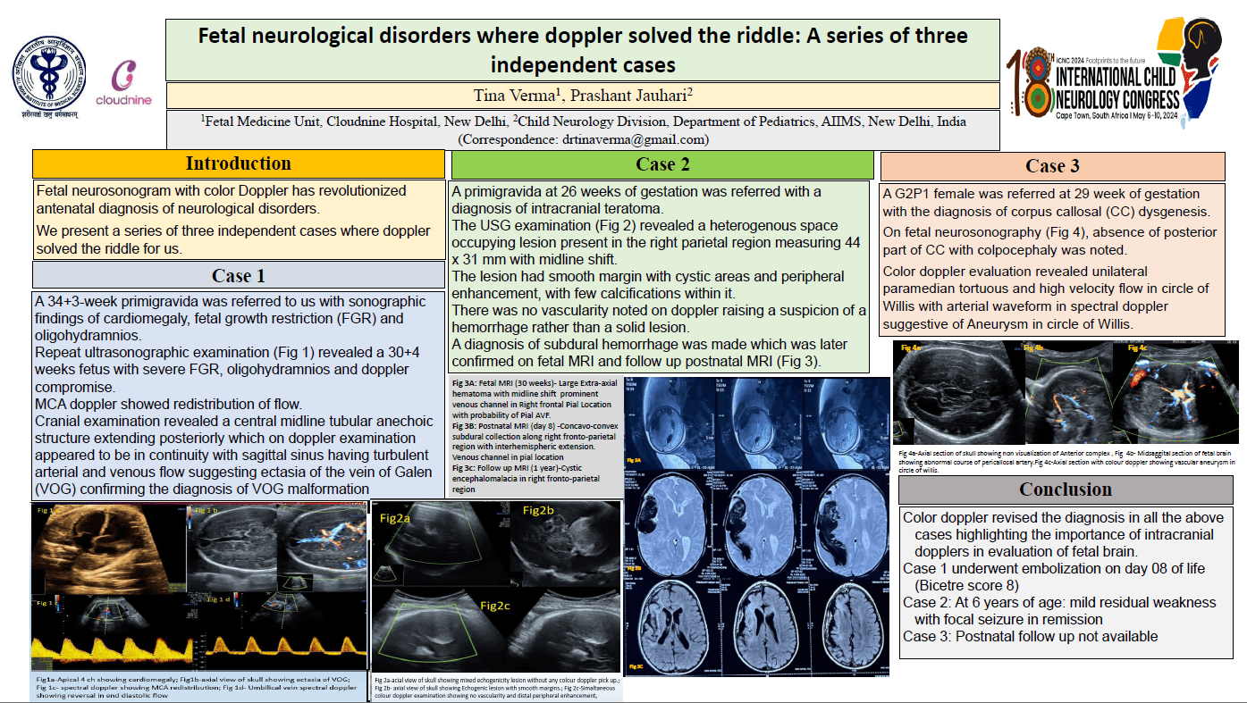
Objectives: We present three independent fetal neurological malformations (on neurosonogram) where color doppler solved the riddle for us. Methods: Case 1: A 34+3-week primigravida was referred to us with sonographic findings of cardiomegaly, fetal growth restriction(FGR) and oligohydramnios. Repeat ultrasonographic examination revealed a 30+4 weeks fetus with severe FGR. Middle cerebral artery doppler showed redistribution of flow. Cranial examination revealed a central midline tubular anechoic structure extending posteriorly which on color doppler appeared to be in continuity with sagittal sinus having turbulent arterial and venous flow within it suggesting ectasia of the vein of Galen (VOG). Case 2- A 26 weeks primigravida was referred with a diagnosis of intracranial teratoma. Repeat neurosonogram revealed a heterogenous space occupying lesion (44 x 31mm)present in the right parietal region with midline shift. The lesion had smooth margin with cystic areas, peripheral enhancement, and few calcifying spots. No vascularity noted on doppler. This raised suspicion of a hemorrhage rather than a solid lesion. A diagnosis of subdural hemorrhage was made, later confirmed on fetal MRI. Case 3- A G2P1 female was referred at 29 week with corpus callosal dysgenesis. On fetal neurosonography, absence of posterior part of corpus callosum with colpocephaly was noted. Color doppler revealed a unilateral paramedian tortuous and high velocity flow in circle of Willis with arterial waveform in spectral doppler suggestive of aberrant arteries/aneurysm. Conclusion: Color doppler revised the diagnosis in all the above cases highlighting the importance of intracranial dopplers in evaluation of fetal brain.
Tina Verma
Cloudnine Hospital, Patparganj
India
Prashant Jauhari
All India Institute of Medical Sciences
India
Prashant Jauhari
All India Institute of Medical Sciences
India