Clinical, Imaging, And Genetic Spectrum Of Polymicrogyria In Indian Children-Single Centre Experience
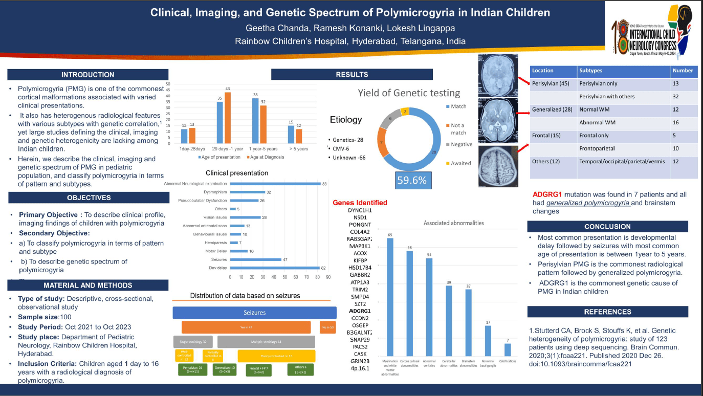
Introduction Polymicrogyria (PMG) is the commonest cortical malformation associated with varied clinical presentations, heterogeneous radiological features, and various subtypes with genetic correlation. However, large studies defining the clinical, imaging, and genetic heterogenicity are lacking among Indian children. We describe the clinical, imaging, and genetic spectrum of PMG, and classify polymicrogyria in terms of pattern and subtypes. Methods Design: Descriptive, cross-sectional, observational study Setting: Department of Neurology, Rainbow Children’s Hospital, India. Results Between October 2021 and October 2023, there were 100 children with polymicrogyria, 59 were boys (59%); 43 children (43%) presented between 1-5 years of age. The commonest manifestations were developmental delay (82 of 100; 82%) and epilepsy (47 of 100, 47%). Among epilepsy, the majority had a single type of seizure (68%) (Table 1). EEG abnormalities were more commonly seen with generalized polymicrogyria. The most common radiological pattern was perisylvian PMG (Figure 1A). The most common associated abnormality was in white matter (65%) (Figure 1B). Unilateral PMG was seen in 23 children and had late presentation and more seizures than bilateral PMG. Only 44 of 100 children underwent genetic testing, of which causative mutation was identified in 28 (63.6%) (Figure 2). Our cohort's most common genetic mutation was ADGRG1 (7 of 28, 25%). Conclusion Perisylvian PMG is the most common pattern in Indian children, with a majority of them having developmental delay and epilepsy. ADGRG1 is the most common genetic cause of PMG in Indian children.
Geetha Chanda
Rainbow childrens Hospital
India
Ramesh Konanki
Rainbow childrens hospital
India
Lokesh Lingappa
Rainbow Childrens Hospital
India
Nihaal Reddy
Rainbow childrens Hospital
India
Ramesh Konanki
Rainbow childrens hospital
India